Siegburg: Joint embolization - new therapy for painful knee prostheses
Embolization instead of painkillers
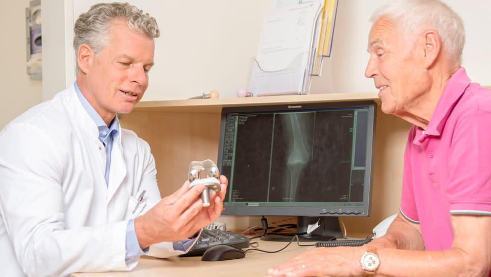
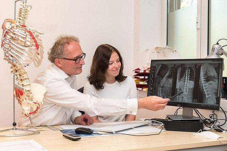
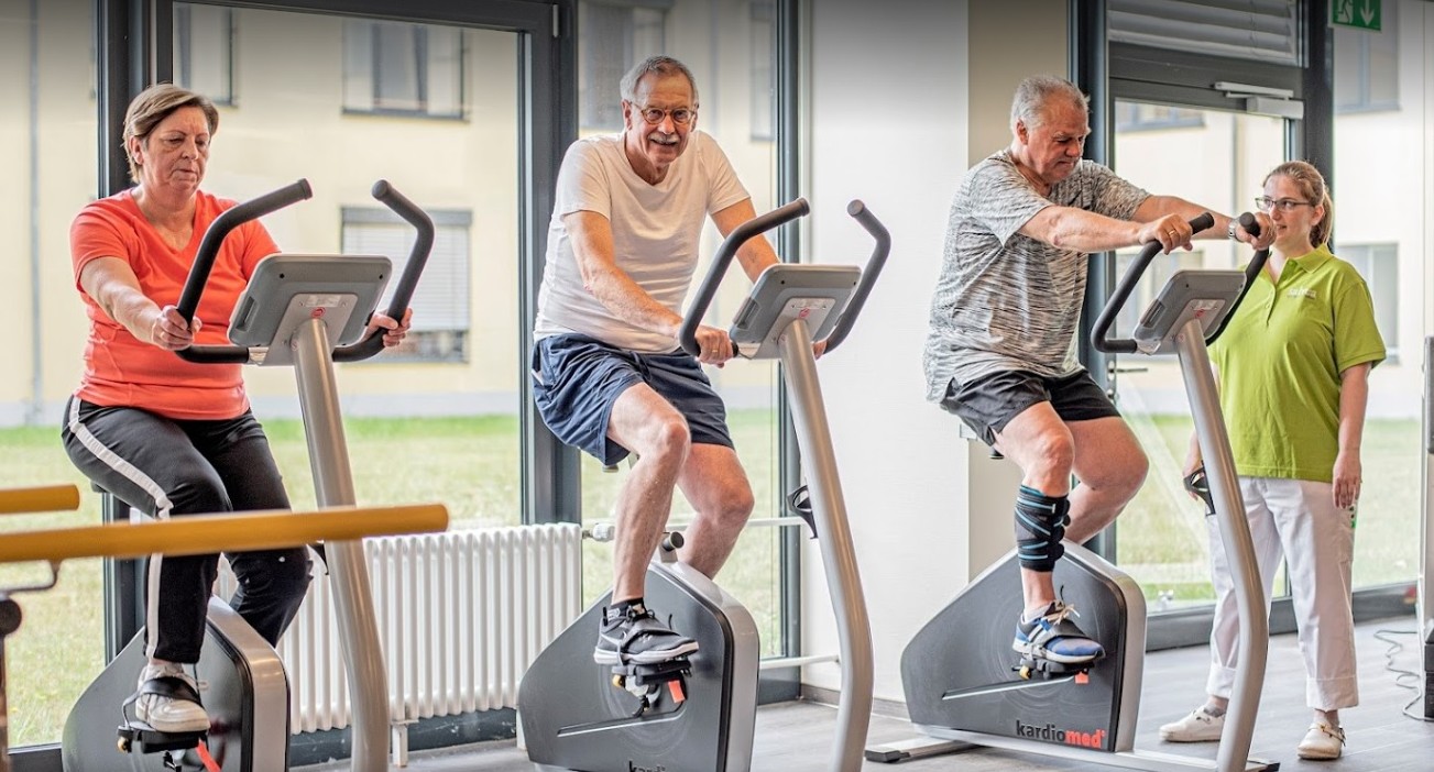
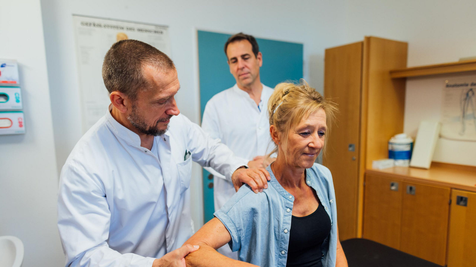
Painkillers often do not respond adequately to chronic knee pain. The joints are permanently irritated and the vessels permanently cause inflammatory reactions and thus pain. The Helios Klinikum Siegburg is therefore using a new, interventional treatment approach: transarterial periarticular embolization, or TAPE for short. "This is known as joint embolization. Under local anaesthetic, a thin catheter is inserted through the groin artery to the affected knee joint as part of an angiography and an antibiotic is injected into the affected, pathologically altered vessels via the catheter," explains Dr. med. Bastian Vloet, Senior Consultant at the Institute of Diagnostic and Interventional Radiology, who performs the intervention in Siegburg.
The antibiotic causes the blood vessels to "stick together" and the nerve endings to die off. "As a result, patients usually experience significant relief from their knee pain very quickly," says Dr. Vloet.
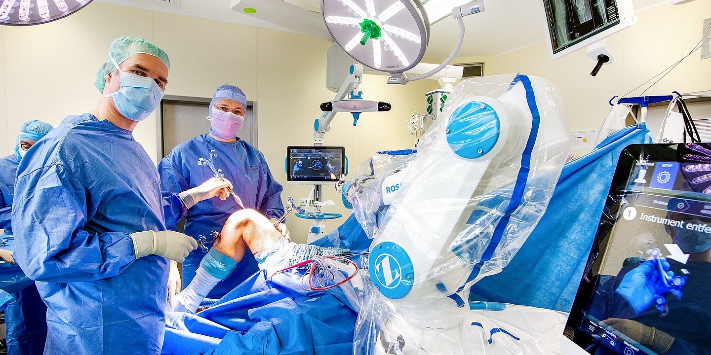
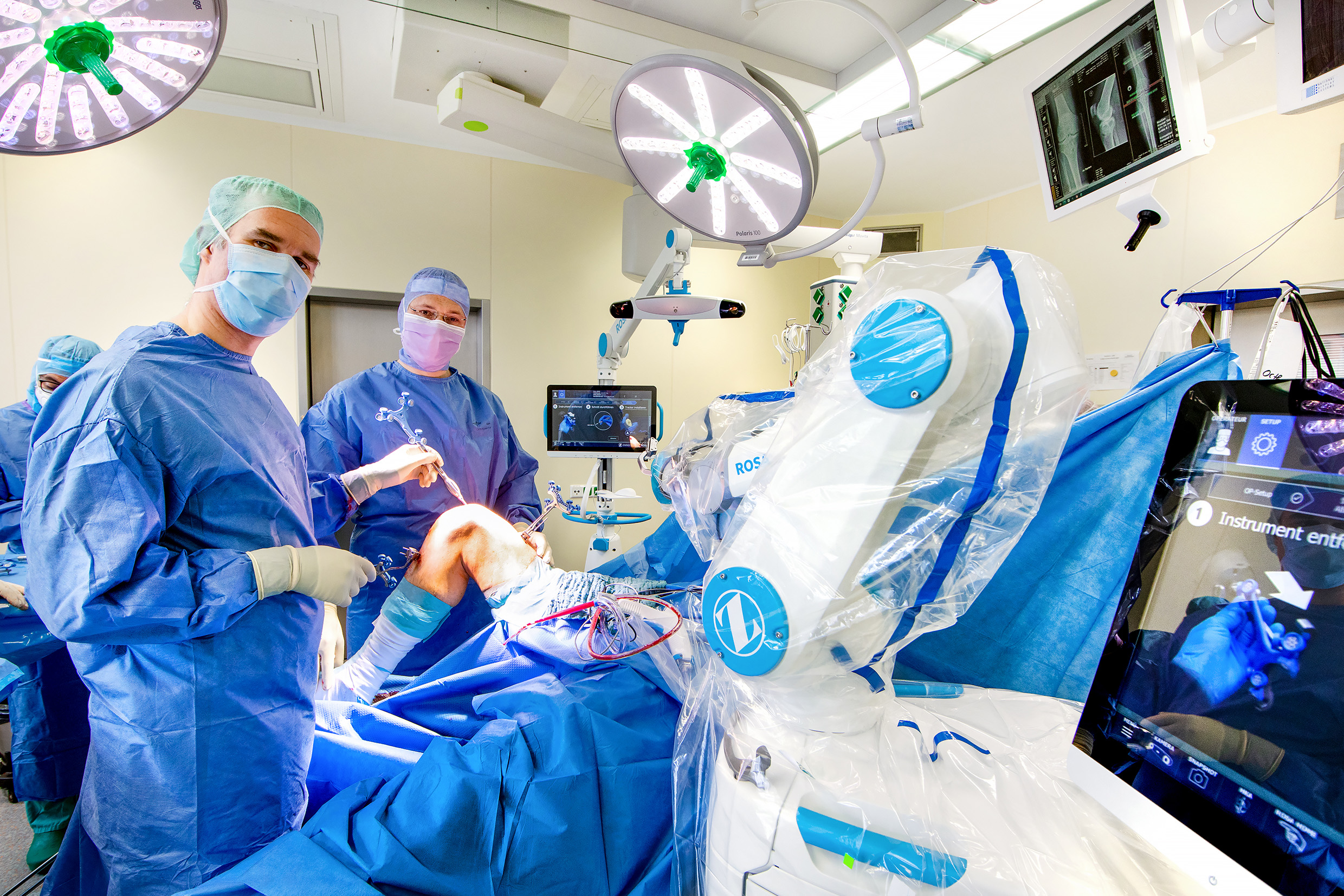
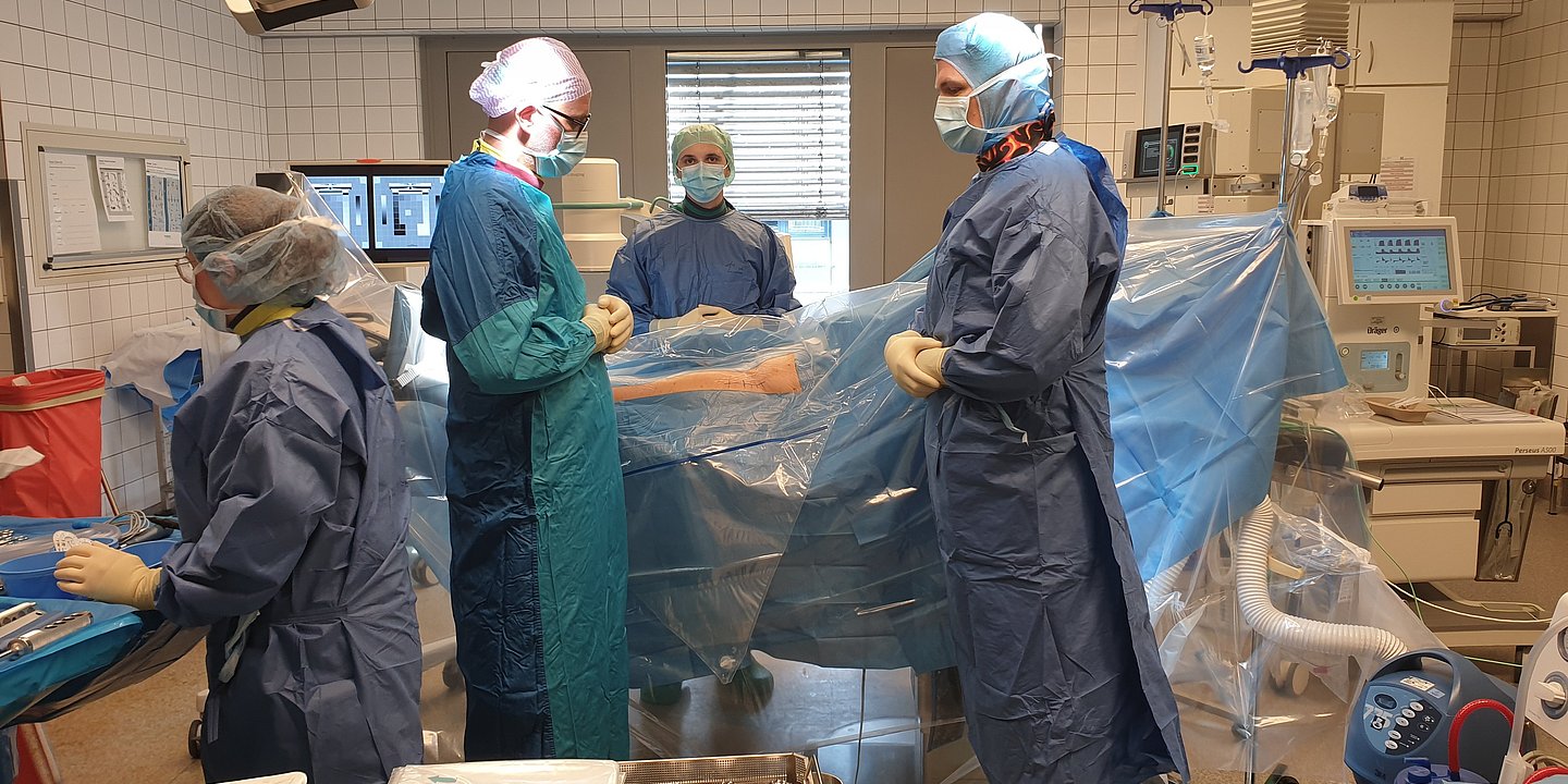
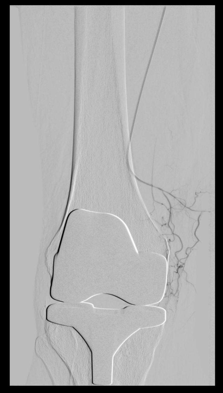
The procedure, which usually takes two hours, takes place in the hospital's catheter laboratory under constant X-ray monitoring. The radiologist uses a contrast medium to visualize the vessels around the knee and can thus check where the antibiotic is to be applied. As the procedure takes place under local anaesthetic, the patients are awake and can communicate with the radiologist. "This is also very important," emphasizes Dr Bastian Vloet. "It allows the patient to tell me exactly whether we have 'hit' the right painful areas."
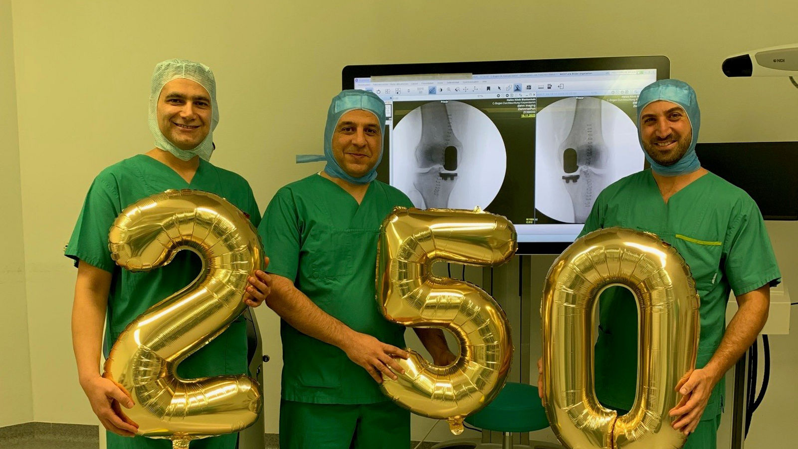
Dr. Vloet and his team perform joint embolizations regularly and very successfully. In addition to painful knee joints, the procedure can also be used for chronic shoulder pain, for example.
"Under local anaesthetic, a thin catheter is inserted through the inguinal artery to the affected knee joint as part of an angiography and an antibiotic is injected via the catheter into the affected, pathologically altered vessels,"
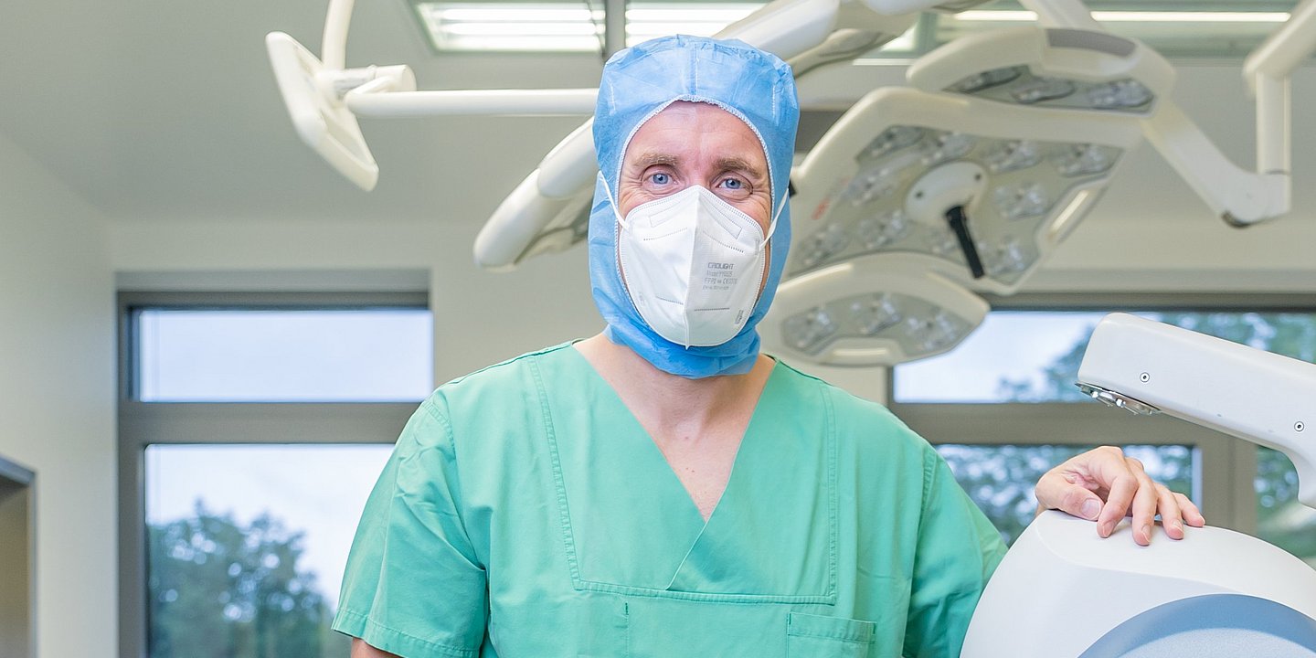
explains Dr. med. Bastian Vloet, EBIR, Senior Consultant and Head of Interventional Radiology and Vascular Medicine.
Read more:
Do you need more information about Helios Hospitals or want to schedule your treatment?